case 14: A Simple Way To Treat A Class I Cavity

Assalammualaikum Wr Wb.. Good Morning!!

Hii.. Good Evening on People from GMT+7 area, hehe.. So, today is february 24th, 2015. I think it is a good time to write down a new case, my recent case which was done at last saturday, february 21st 2015. So,  welcome to SekilasKonservasiGigi.Com, conservative dentistry at a glance.. a mini story of CeritaPasienRio.Com.. 🙂 this site will be delivered in english, so, you would not need any google translator, hehe..

in this midweek, i will present to you my restoration on class I cavity, yup! still in molar. for some dentists might feel it is a very common case, but for me as the other one might think that it is a basic but challenging. hehe.. at least dentistry is very challenging now a days huh.. hehe..

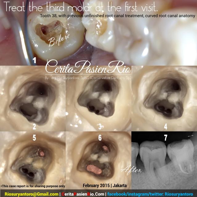
i got this case last saturday, a patient who want to go to australia to continue the her study, she had her full dental health checked before going to Aussie.. So, i found an interesting class I cavity here in tooth 36. this molar has many deep fissures, and a deep caries progression.

I restored the cavity using a composite resin, SDR as a dentin replacement and Ceram.X one as upper part of restoration. the time lapse was 30 minutes including a polishing and finishing time. Oh, i used a single shade, no tint or color modifier.. so, here it is.. 🙂

dokter gigi rio suryantoro spesialis konservasi gigi resin komposit kelas 1


Picture Information:

1. (upper left) First appearance, yup.. as you see, the caries location was at occlusal surface or we called it class I cavity. this tooth anatomy had many deep fissures which also had a caries.. 

2. (upper right) tooth appearance after caries removal and cavity preparation. ready to fill it with composite resin

3. (middle left) ah, this was the appearance after filling with composite resin packable and SDR as dentin replacement. ready to finish and polish..

4. (middle right) the tooth checked for occlusion and articulation behaviour.

5. (bottom) FINAL RESULT.

Yup, that’s for my 14th story in SekilasKonservasiGigi.Com, For more stories please visit CeritaPasienRio.Com or browse my fb at facebook.com/RioSuryantoro and instagram.com/Riosuryantoro

don’t forget to click the follow botton on your rigth side to get the latest updates from my site..

see you on my next dental story..

thank You Very Much,

keep UP the GOOD WORK!!

keep on Sharing,

Wassalammualaikum Wr Wb..

-drg. Rio Suryantoro, Sp.KG-

M e e t   m e   a t :

socmed publication

Posted in Uncategorized | Leave a comment

case 13: Treat the third molar at the first visit

Assalammualaikum Wr Wb.. Good Morning!!

Today, february 12th 2015, is a good day to write down a new case report which was done in february 10th 2015, yup! fresh case two days ago.. 🙂 welcome to SekilasKonservasiGigi.Com, conservative dentistry at a glance.. a mini story of CeritaPasienRio.Com.. 🙂

hmm… today i got a very sad news that there were a shooting accident to three muslims in chapel hill, i wish for their spirit. And also this week is a very rainy season in Jakarta, many floods happen every where. (even, i could not go to my dental practice last monday), i wish for a better jakarta.. 🙂

ok.. now is the turn to share my case. as i mentioned before in my tittle, this case will tell the story about third molar. The patient came to me with mild to moderate pain, he/she had went to many dentists and got many advices wether he/she got him/her tooth removed or treated. the tooth happened to get a dental treatment before by my patient’s previous dentist, when i checked the tooth, it was a GIC and had broken structure already. after got the informed consent by the patient, i started to treat the tooth. the first obstacle was the location of the tooth, yes it is a third molar with a small mouth opening and limited working area. Second is the anatomy of the root canals, oh.. they are curved canal. here is the case.. 🙂 sapti psa 38 bengkok doker gigi rio suryantoro spesialis konservasi gigi perawatan saluran akar one visi

Picture Information:

1. First appearance, take a look at the restoration which already destructed, and the caries progression into the pulp. i suspected that the previous operator had tried to do an open access

2. after access opening, i found 2 out of 3 canals. but the 10 k-file had reached the mesiobuccal successfully.

3. take a look at the mesio-buccal canal which already opened after doing the mesiolingual and distal canals.

4. finished root canal cleaning and shaping.

5. filling the distal canal

6. finished filling the 3 canals.

7. radiographic image after obturation and placement of the coronal seal (also as a canal anchorage).

the files i used in here were Protaper, with sodium hypochlorite 5,25% as irrigation material. 

Yup, that’s for my 13th story in SekilasKonservasiGigi.Com, For more stories please visit CeritaPasienRio.Com or browse my fb at facebook.com/RioSuryantoro and instagram.com/Riosuryantoro

see you on my next dental story..

thank You Very Much,

keep UP the GOOD WORK!!

keep on Sharing,

Wassalammualaikum Wr Wb..

-drg. Rio Suryantoro, Sp.KG-

M e e t   m e   a t :

socmed publication

Posted in Uncategorized | Leave a comment